


Deforming arthrosis of the knee joint is a polyetiological disease. This means that there are a lot of reasons for its development. In some cases, when the most dominant cause can be allocated, gonarthrosis is called secondary. In the case when a clear cause is not determined, the diagnosis of primary or idiopathic arthrosis of the knee joint is established.
- deforming osteoarthrosis of the medial part of the femoral-bore joint;
- deforming osteoarthrosis of the lateral part of the femoral-bore joint;
- deforming arthrosis of the femoral-delicious joint.
Normally, the destruction of the joint cartilage occurs in the process of physiological wilting of the whole organism, that is, during aging. The pathological destruction of the cartilage is considered when it occurs ahead of time or more intense pace. The average age, at which on a completely legitimate basis, the first signs of cartilage degeneration can manifest itself is a period of 40 to 50 years. With deforming arthrosis, the disease debuts in childhood with the first manifestations of 16 - 18 years, and in some cases even earlier. However, this is not a reason to despair.
The mechanism of the development of the disease is a vicious circle in which the final links launch the initial and so on to infinity. However, each round of this circle exacerbates the condition of the cartilage and leads to the progression of the disease and so on the chain. In the case of primary gonarthrosis, the reason that launches a vicious circle is unknown. Nevertheless, its subsequent links are carefully studied with the aim of influencing them and slowing down the progression of the disease.
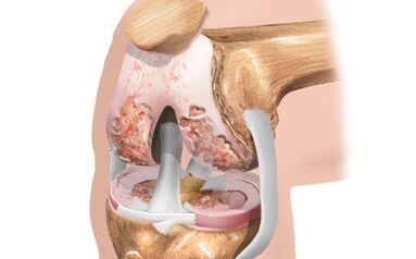
Deforming arthrosis develops approximately as follows. Daily joint cartilage of the knee joint experience thousands of shocks that they are forced to depreciate in order not to harm the more tender structures of the human body, such as internal organs and brain. Over time, due to the data of concussions, microscopic cracks are formed in the cloudy layer, which also after a certain period of time are filled with synovial fluid and turn into microdists. Neighboring microdists have a tendency to unite and formation of larger cysts.
Increasing in the size of the cysts of the corneal space gradually begin to squeeze blood capillaries that feed the cartilage tissue from the side of the bone. The supply of it with oxygen and substances necessary to maintain vital activity, worsens, which leads to a more slow synthesis of type 2 collagen.
Carriage of cartilage leads to two negative consequences. Firstly, it leads to a deterioration in the depreciation properties and a more intensive formation of new microcracks in the subsidiary layer. Secondly, due to compression of the cartilage, its density increases, which adversely affects the second mechanism of its nutrition-through the diffusion of synovial fluid into the thickness of the cartilage tissue.
Nevertheless, on the scale of the whole organism, the destruction of the articular cartilage does not go unnoticed. As a compensatory reaction in the focus of washing cartilage, the activity of chondroblasts - young cells synthesizing new cartilage tissue increases. However, this compensatory mechanism is imperfect, and its imperfection lies in the fact that the bulk of the cartilage tissue is formed not at the place of the greatest destruction of the cartilage, but where the cartilage does not experience loads.
As a result, the cone -shaped growths of cartilage are formed along the edges of the joint - chondroophytes. These chondroophytes are clinically not manifested until the processes of ossification begins in them. Okreteen, chondroophytes harden and turn into, which are called spikes in common people. As a rule, the appearance of spikes is always accompanied by the occurrence of pain and the development of inflammation in the joint. This is due to the fact that osteophytes during the movement of the joint touch the cartilage tissue and the synovial shell, thereby mechanically damaging it.
As a result, each complication of deforming arthrosis leads to the acceleration of the progression of pathological changes in cartilage. However, knowing the mechanism of development of gonartrosis, one can successfully affect some of its links in order to slow down its current and improve a long -term forecast.
Secondary gonarthrosis differs from the primary one in that the main reason is known, which launched a vicious circle of destruction of the joint cartilage. The further course of the disease occurs in the same way as with primary gonarthrosis, with the peculiarity that the disease is constantly aggravated by the effect of negative factors associated with the underlying disease. For this reason, the course of secondary arthrosis of the knee joint, as a rule, is more aggressive.
- injuries (acute and chronic);
- congenital varus or valgus deformation of the lower extremities;
- congenital shortening of one of the lower extremities;
- hypermobility syndrome of the knee joint;
- congenital dysplasia of the knee joint;
- chondrocalcinosis;
- osteomyelitis;
- rheumatoid arthritis;
- acromegaly;
- diabetes mellitus;
- obesity;
- hypothyroidism;
- Fraud, etc.
Post -traumatic deforming arthrosis is divided into acute and chronic. The acute form of the disease develops after one serious injury, more often -
, which occurs or partially extends to the articular part of the bone. The chronic form of the disease develops for a longer time and is associated, as a rule, with frequent and slight injury to the joint. Such conditions are created by builders, road workers, movers, etc.
In acute gonarthrosis, the mechanism of the disease is associated with severe inflammatory changes in the joint cavity, namely with lymphostasis, increased pressure in the joint cavity, and a change in the composition of the synovial fluid. Excessive acceleration of the growth of new cartilage fabric leads to deformation of the articular surface at the fracture site and the growth of osteophytes.
In chronic gonartrosis, a severe inflammatory process is not observed, however, the frequent and intensive load on the cartilage tissue leads to its rapid compression, the formation of microcracks and the deterioration of cartilage supply with nutrients both from the side of the bone and from the joint gap.
People with this pathology can be found quite often. Its essence is to change the shape of the legs. With varior deformation, the legs are arched outward in a horizontal plane. In other words, between the patient’s legs, the space is more than in healthy people. With valgus deformation, the legs have a X-shaped shape when the knees are in contact with each other. Both pathologies can be both genetically programmed and developing throughout life due to fractures of the lower extremities.
In both cases, the load on one of the sides of the knee joint increases, with varus deformation - on the lateral sides, and with valgus deformation - on the medial sides. Due to the fact that the same weight of the patient presses on a smaller area, premature washing of the cartilage occurs, accompanied by inflammation, pain and morning stiffness.
Congenital shortening of one of the legs is a consequence of anomalies or can develop a few years after birth as a consequence of the birth injury. As in the previous case, an uneven distribution of weight occurs, and the normal leg takes on a large load. As a result, the joint cartilage of the knee joint of a healthy leg undergoes structural changes that lead to deforming arthrosis.
This pathological condition is not a disease, but it may well lead to it. This syndrome means excessive mobility of the ligamentous and consistent apparatus, in which the joint movement of the joints within normal axes can significantly increase. Such patients almost never suspect that they have such a feature, since they live with it all their lives and believe that other people also function in the same way.
A sign of hypersmors of the knee joint is the formation of a stupid angle between the front surfaces of the thigh and the lower leg with maximum straightening of the leg. In other words, the knees are bending as it were, and the legs take an ardent shape. Also, such patients can easily reach the forearm with his thumb, reach their heads to the legs and, in principle, have congenital flexibility.
Symptoms of arthrosis of the knee joint
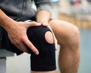
At the initial stage of development, pathology is manifested by pain in the knee, moderately expressed and arising while in motion, when moving along the steps.
An unpleasant symptom may appear if a person spends a lot of time standing or tries to rise after being in a sitting position for a long time.
At rest, health usually improves.
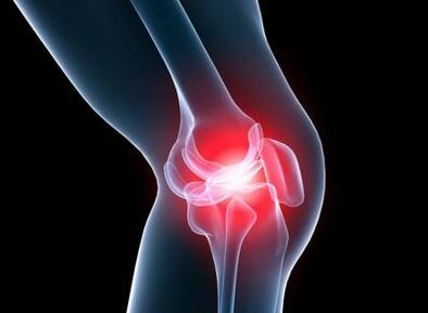
Sharp intense severe pain arises spontaneously.
Most patients previously had prolonged discomfort during physical activity and when walking. In this case, growing pain may be the main sign of the development of gonarthrosis.
The disease develops gradually, for several months or years, when not yet visible deformation and severe pain. But during this period, discomfort in the knees, which occurs from time to time.
Remember, the earlier you consult a doctor, the easier and more successfully the treatment will pass.
Do not delay a visit to a specialist, waiting for irreversible consequences. Take measures as soon as you notice the symptoms of the disease.
The obvious signs of arthrosis of the knee joint begin to manifest as the structure of the cartilage shells, a decrease in the production of synovial fluid and damage to the joint bag. At the initial stage of the increase in pathological changes, as a rule, there is no pronounced symptoms, but at the same time a slight stiffness may be present in the morning.
When pronounced and various symptoms appear, arthrosis, as a rule, is already in the late stage of their development. At this time, there is already serious damage to the structures of the knee joint, so the disease goes into the acute phase. The characteristic manifestations of the acute period of development of arthrosis include:
- increased pain;
- Change of gait;
- lameness;
- crunch when moving;
- swelling of soft tissues;
- an increase in the knee due to the accumulation of liquid in it;
- Limiting mobility in the joint.
When knee arthrosis develops, the symptoms can grow long enough, but when the disease is transition to the last 3 stage of manifestation of a disorder of the joint, the quality of life significantly reduces the quality of life.
- The synovial shell against the background of damage to the articular surfaces begins to become inflamed, which leads to a violation of the mobility of the entire joint.
- Any movement with a damaged joint can be very painful.
- On palpation, a significant increase in local body temperature is noted.
- As a rule, the effective treatment of arthrosis of the knee joint with conservative methods is possible only at the early stage of the development of the disease.
- Thus, when signs of the disease appear, consult a doctor for consultation.
In the development of such a condition as the arthrosis of the knee, the symptoms and treatment are interconnected, since if at the early stages of the development of the disease the articular surfaces can still be completely restored and improved by local metabolism, then in the later stages the drug treatment of arthrosis of the knee joint can often not have a positive effect, since cartilage tissues are so thin that bone structures are exposed.
Arthrosis of the 1st degree proceeds almost without visible symptoms. This phase of development is characterized by:
- fatigue in the legs;
- A slight decrease in mobility, which is usually observed immediately after sleep.
Pain symptoms, if and occur, are manifested to a slight degree. At this moment, the arthrosis of the knee looks in the x -ray in the form of small bumps on the cartilage tissue and the surface of the bones.
With arthrosis of the knee joint of the 2nd degree, the symptoms are more pronounced. Pain arises already from the minimum load or immediately after it. In the affected part of the leg, the pain is caused by almost any movement. After a fairly long rest, it usually passes completely. However, the next physical actions immediately cause pain.
At approximately the second stage of the development of the disease, the pain sensations are added:
- crunch in the knee joint during movements;
- a reduced opportunity to bastard the leg normally in the knee;
- change of joint bones;
- Progressive synovitis.
A rough arthrose crunch of joints, as a rule, is first barely audible, but with the course of the disease it becomes very loud and distinct. When trying to bend the leg in the knee, a sharp pain occurs. In some cases, this is possible only to a corner of 90 degrees, and then with difficulty and overcoming pain. The change in the shape of the joint also becomes obvious, which is also aggravated by the accumulation of pathological fluid in it.
The characteristic features of the 3rd degree of arthrosis are severe pains that are independent of the amount, the intensity of physical activity. The joint bothers a person even at night, this causes significant inconvenience.
The radiograph can show global changes in cartilage, joint surface, non -characteristic growths. O-shaped or X-shaped curvature leads a person to disability. These are the consequences that the cartilage tissue has already worn out and bone tissue went into the "move".
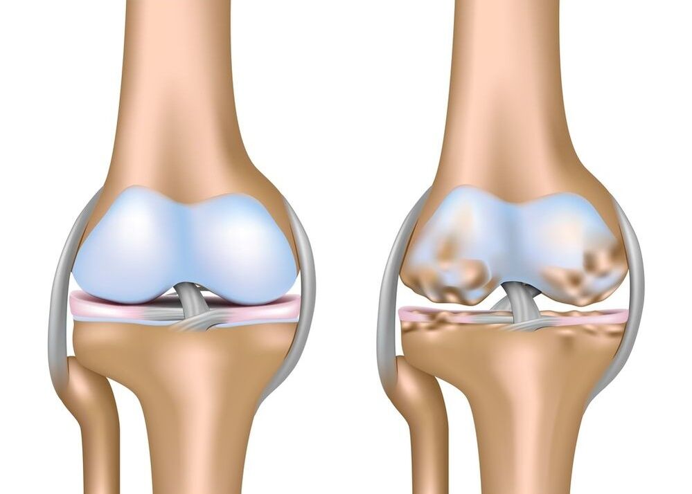
Gonarthrosis is a disease of degenerative-dystrophic nature, in which the destruction of cartilage occurs and the joint is deformed. Signs of the disease are severe pain, deformation of the limbs, an uneven distribution of load on the bone-muscular system, the development of complications and a significant decrease in mobility up to disability of the patient.